3D Printer Builds Artificial Blood Vessels
The goal is to custom-build tissues and organs for transplant, using the patient's own cells and 3D medical printers.


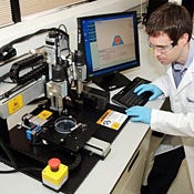
Invetech Orgonovo 3D Medical Printer
(click image for larger view)
Invetech Orgonovo 3D Medical Printer
A San Diego company is developing technology to "print" artificial blood vessels for transplant. The initial goal: Create an arterial graft for use in coronary bypass surgery.
The long-term goal is to solve problems in medical therapy that can't be solved otherwise, especially in organ transplants, where tens of thousands of people are waiting for donated organs, said Keith Murphy, CEO of the company, Organovo.
Invetech, a design and contract manufacturing company with offices in Australia and San Diego, built Orgonovo's first 3D medical printer, in conjunction with Organovo.
"Building human organs cell-by-cell was considered science fiction not that long ago," said Fred Davis, president of Invetech, in a statement. "Through this clever combination of technology and science we have helped Organovo develop an instrument that will improve people’s lives, making the regenerative medicine that Organovo provides accessible to people around the world.”
Murphy said in a statement, "Scientists and engineers can use the 3D bio printers to enable placing cells of almost any type into a desired pattern in 3D.” He added, "Researchers can place liver cells on a preformed scaffold, support kidney cells with a co-printed scaffold, or form adjacent layers of epithelial and stromal soft tissue that grow into a mature tooth. Ultimately the idea would be for surgeons to have tissue-on-demand for various uses, and the best way to do that is get a number of bio-printers into the hands of researchers and give them the ability to make three dimensional tissues on demand.”
The technology works by using a robot to lay down cells in precise positions in three dimensions, accurate to within 20 microns. "It's similar to the way a laser printer prints by putting solid particles in place," Murphy told InformationWeek. The 3D medical printer puts down objects on 2D layers, one on top of the other. The particles used in the construction are made up of stem cells, formed into tiny spheres and cylinders.
The stem cells are available for research purposes from companies including Life Technologies and Invitrogen. When the device is used for treatment, cells will come from the patient, such as bone marrow, or fatty adipose tissues, where stem cells can be harvested. "Because they come from the patient, there's no risk of having a rejection," Murphy said. These are adult stem cells, not the fetal stem cells that have been politically controversial.
Researchers take a cross-section picture of the object they want to build, such as an artery. "We use that as a map to paint by numbers," he said.
Objects take about an hour to build, and then the cells fuse together on their own in the course of 24-48 hours, locking the object in shape.
3D printers are an emerging technology with a wide variety of applications. Like Organovo's equipment, they build 3D objects by laying down two-dimensional layers one on top of the other. They typically use plaster, cornstarch or resins to create objects, and are most often used in rapid prototoyping, for footwear, jewelry, industrial design, architecture, automotive, aerospace, dental, and medical industries.
Organovo is not alone finding medical applications for 3D printing; researches at the University of Tokyo hospital and venture company Next 21 are using 3D printers to create artificial bones for reconstructive surgery.
Organovo is starting simple, building blood vessels at first, including an arterial structure meant for use in coronary bypass surgery. That involves creating an object with three different cell types in it: Endothelium cells on the inside, smooth muscle in the middle, and an exterior layer of fibroblasts, which are similar to skin cells.
The arterial segments are 5-20 centimeters long, with an interior diameter of 0.5 to 5 mm. Arteries with larger interior diameters can be built with Teflon or Dacron, but the smaller diameters clot when built using synthetic materials.
A next step will be to make a branched structure of blood vessels, which will allow putting the artificially grown vasculature inside living tissue. "Sure, I can make a one-inch cube of liver tissue now, but they'll all die because they can't get nutrients," Murphy said. With a branched structure, scientists can make larger, thicker pieces of tissue and keep them alive with blood vessels inside.
The printed blood vessels should be in clinical trials in three to five years, with thicker, and more complex types of tissues further out. In the long run, researchers hope to build a kidney or heart or other organ. "That's certainly within the long-term capabilities. But we're focused as a company on what works short-term from a commercial standpoint, and that allows us to move forward and consider longer-term things," Murphy said.
"The printer fits inside a standard biosafety cabinet for sterile use," Invetech said. "It includes two print heads, one for placing human cells, and the other for placing a hydrogel, scaffold, or support matrix. One of the most complex challenges in the development of the printer was being able to repeatedly position the capillary tip, attached to the print head, to within microns. This was essential to ensure that the cells are placed in exactly the right position. Invetech developed a computer controlled, laser-based calibration system to achieve the required repeatability."
Invetech plans to ship multiple 3D bio-printers to Organovo in 2010 and 2011. Organovo will place the printers globally with researchers in centers for excellence for medical research.
Orgonovo was co-founded by Gabor Forgacs, a professor of biophysics at the University of Missouri, who studied chicken embryo development to determine how cells in the embryo move around to form tissues--for example, a wing--from a small number of cells in the beginning. "That study enabled him to get a good handle on how clumps of cells grow into tissues," Murphy said.
Forgacs' research was funded by a $5 million grant from the National Science Foundation, and the technology is licensed by by Orgonovo from the University of Missouri. The company is angel-funded, and will be seeking venture funding in 2010.
About the Author
You May Also Like


